5.4 ANATOMY OF ROOT, STEM AND LEAF :
5.4.1 Anatomy ofDicot Root
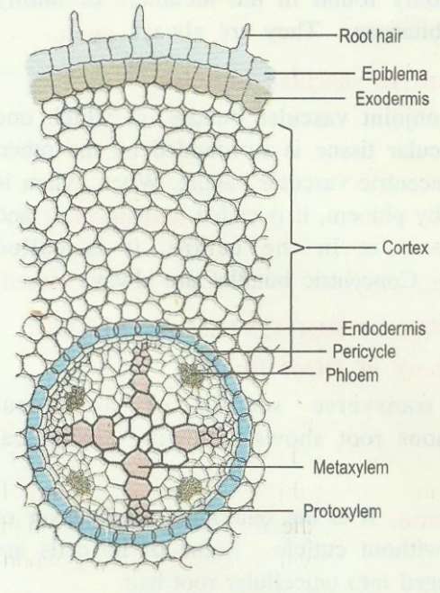
The transverse section of a typical dicotyledonous root shows following anatomical features.
1. Epiblema:
It is the outermost single layer of cells without cuticle.
Some of its cells are prolonged into unicellular root hair.
2. Cortex:
The cortex consists of several layers of typical parenchymatous cells.
As epiblema dies off outer layer of cortex become cutinized and is called exodermis.
The cortical cells store food (mostly in the form of starch) and water.
3. Endodermis:
The innermost layer of cortex is called endodermis.
Its cells are barrelshaped and their radial walls.
It bears Casparian strip or Casparian bands composed of suberin.
Near the protoxylem there are unthickened passage cells.
4. Pericycle:
Below the endodermis a single layer of parenchymatous pericycle is present which bounds the stele or vascular cylinder.
5. Stele:
It consists of 2 to 6 radial vascular bundles.
(The xylem and phloem are arranged alternately on different radii).
Xylem is exarch and consists of tracheids, vessels, parenchyma and sclerenchyma.
The phloem consists of sieve tubes, companion cells and phloem parenchyma.
Based on the number of groups of xylem and phloem, the stele may be diarch to hexarch.
6. Connective tissue:
A parenchymatous connective tissue or conjunctive tissue is present between xylem and phloem.
7. Pith:
The central part of stele or vascular cylinder is called pith.
It is narrow and made up of parenchymatous cells, with or without intercellular spaces.
At later stage, a cambium ring develops between xylem and pho1em which causes secondary growth in thickness.

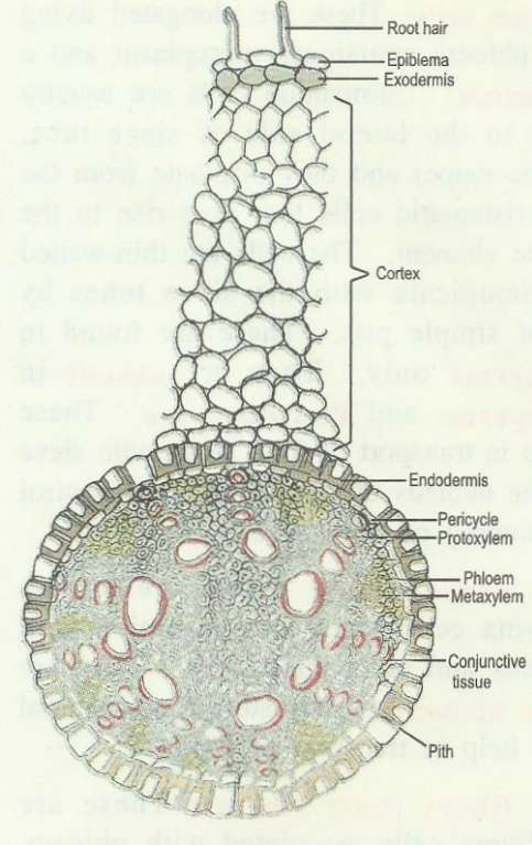
Anatomy of a monocot root
It resembles that of a dicot root in its basic plan.
However, it possesses more than six xylem bundles (polyarch condition).
Pith is large and welldeveloped.
Secondary growth is absent.

