8.8 Anatomy of Root, Stem and Leaf :
A. Anatomy of Dicot Root :
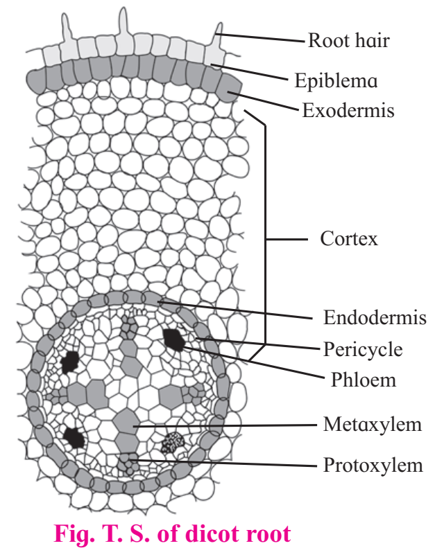
The transverse section of a typical dicotyledonous root shows following anatomical features.
The outermost single layer of cells without cuticle is Epiblema. Some of its cells are prolonged into unicellular root hair.
Next to it on inner side is the Cortex which consists of several layers of typical parenchymatous cells. After the death of epiblema, outer layer of cortex become cutinized and is called Exodermis. The cortical cells store food and water.
The innermost layer of cortex is called Endodermis. The cells are barrel-shaped and their radial walls bear Casparian strip or Casparian bands composed of suberin. Near the protoxylem, there are unthickened passage cells. A single layer of parenchymatous
Pericycle is present just below endodermis which bounds the stele or vascular cylinder.
Stele consists of 2 to 6 radial vascular bundles. Xylem is exarch. Based on the number of groups of xylem and phloem, the stele may be diarch to hexarch.
A parenchymatous connective tissue or conjunction tissue is present between xylem and phloem.
The central part of stele or vascular cylinder is called Pith. It is narrow and made up of parenchymatous cells, with or without intercellular spaces.

At later stage, a cambium ring develops between xylem and phloem which causes secondary growth in thickness.
B. Anatomy of monocot root :
It resembles that of a dicot root in its basic plan. However, it possesses more than six xylem bundles (polyarch condition). Pith is large and well-developed. Secondary growth is absent.

C. Anatomy of Dicot Stem (Sunflower) :
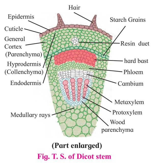
A transverse section of dicot stem shows the following structures :
Epidermis is single, outermost layer with multicellular having outgrowth called trichomes. A layer of cuticle is usually present towards the outer surface of epidermis.
Cortex is situated inner to the epidermis and is usually differentiated into three regions namely, hypodermis, general cortex and endodermis.
Hypodermis is situated just below the epidermis and is made of 3-5 layers of collenchymatous cells. Intercellular spaces are absent.
General cortex is made up of several layers of large parenchymatous cells with intercellular spaces.
Endodermis is an innermost layer of cortex which is made up of barrel shaped cells. It is also called starch sheath.

Stele is the central core of tissues differentiated into pericycle, vascular bundles and pith.
Pericycle is the outermost layer of vascular system situated between the endodermis and vascular bundles.
In sunflower, it is multilayered sclerenchymatous in the region of vascular bundles and it is called hard bast.
Vascular bundles are conjoint, collateral, open, and are arranged in a ring.
Each one is composed of xylem, phloem and cambium.
Xylem is endarch.
A strip of cambium is present between xylem and phloem.
Pith is situated in the center of the young stem and is made up of large-sized parenchymatous cells with conspicuous intercellular spaces.
D. Anatomy of Monocot Stem :
It differs from dicot.
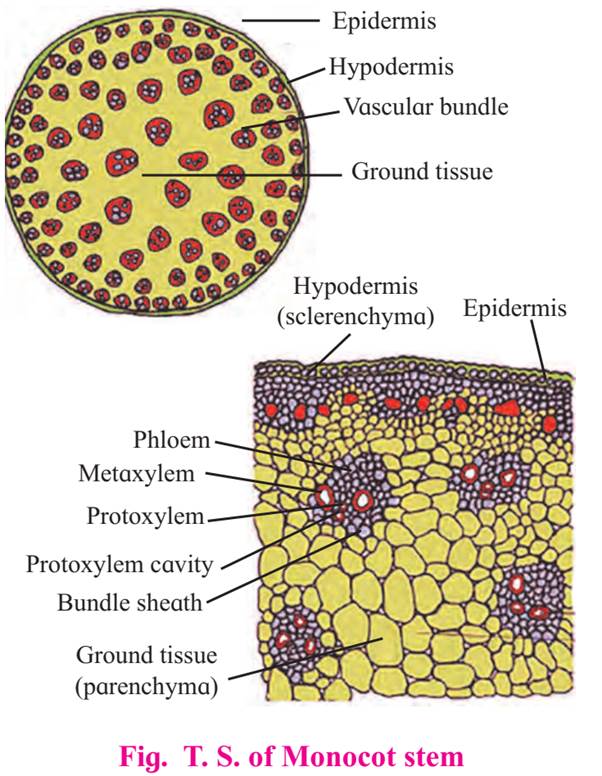
Epidermis is without trichomes and the hypodermis is sclerenchymatous.
Vascular bundles are numerous and are scattered in ground tissue.
Each vascular bundle is surrounded by a sclerenchymatous bundle sheath.
Vascular bundles are conjoint, collateral and closed (without cambium).
Xylem is endarch and shows lysigenous cavity.
Pith is absent.
Secondary growth is also absent.

E. Anatomy of Leaf :
Dorsiventral Leaf is very common in dicotyledonous plants where the mesophyll tissue is differentiated into palisade and spongy parenchyma. The leaves are commonly horizontal in orientation with distinct upper and lower surfaces. The upper surface which faces the sun is darker than the lower surface.
T. S. of Typical dicot leaf :
Upper epidermis consists of a single layer of tightly packed rectangular, barrel shaped, parenchymatous cells which are devoid of chloroplast.
A distinct layer of cuticle lies on the outside of the epidermis. Stomata are generally absent.
Between upper and lower epidermis, there is chloroplast-containing photosynthetic tissue called Mesophyll.
Mesophyll is differentiated into palisade and spongy tissue.
Palisade parenchyma is present below upper epidermis and consists of closely packed elongated cells. The cells contain abundant chloroplasts and help in photosynthesis.
Spongy parenchyma is present below palisade tissue and consists of loosely arranged irregularly shaped cells with intercellular spaces. The spongy parenchyma cells contain chloroplast and are in contact with atmosphere through stomata.

Vascular system is made up of a number of vascular bundles of varying size depending upon the venation.
Each one is surrounded by a thin layer of parenchymatous cells called bundle sheath.
Vascular bundles are closed and xylem lies towards upper epidermis and phloem towards lower epidermis. Cambium is absent hence no secondary growth in the leaf.
Lower epidermis consists of a single layer of compactly arranged rectangular, parenchymatous cells.
A thin layer of cuticle is also present. The lower epidermis contains a large number of microscopic pores called stomata. There is an air-space called substomatal chamber at each stoma.
F. Isobilateral Leaf :
In this leaf both the surfaces are equally illuminated as both the surface can face the sun, and show similar structure.
The two surfaces are equally green.
Generally monocotyledonous plants have isobilateral leaves.
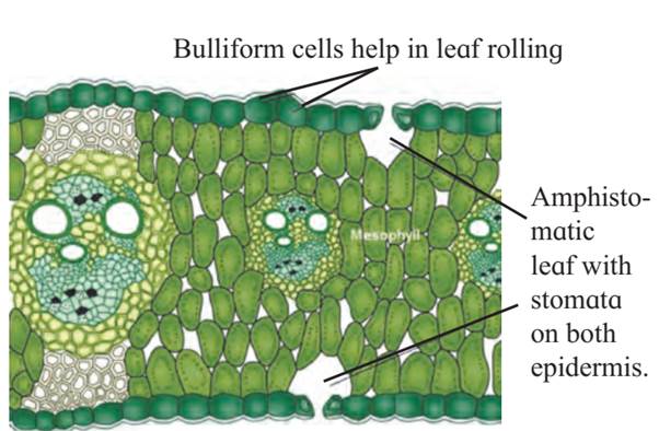
A typical monocot leaf :
It resembles a dicot leaf in its anatomical structure. However, it shows stomata on both the surfaces and mesophyll is not differentiated into palisade and spongy tissue. It has parallel veins. These are conjoint, collateral and closed.

