Structure of Anther
Androecium is the male reproductive floral whorl, having definite number of individual members called stamens.
A stamen has filament, connective and anther.
Anther is the fertile or genesious part of a stamen and usually consists of two anther-lobes (dithecous).
Anther is connected to the filament by sterile connective.
Internally a dithecous anther is tetra-locular i.e. it consists of four chambers called microsporangia or pollen sacs or pollen chambers.
Microsporogenesis i. e. formation of microspores takes place inside the microsporangia or pollen sacs.
T. S. of anther
It shows following structure:
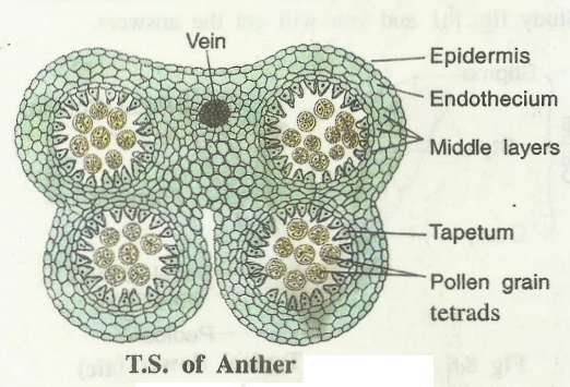
Anther wall:
A mature anther shows wall consisting of following four layers.
Epidermis:
It is the outermost common wall layer of anther which consists of flattened cells. It is protective in function.
Endothecium:
It is internal to the epidermis, common for the four pollen sacs and consists of single layer of cells.
The cells of endothecium show characteristic fibrous thickenings of callose.
Cells of endothecium situated in the shallow groove between two microsporangia remain thin walled and represent the line of dehiscence.
Fibrous thickenings and hygroscopic nature of endothecium cells help in the dehiscence of anther at maturity.
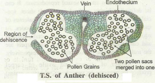
Middle layers:
Internal to endothecium, 1 to 3 layers of parenchyma cells are present surrounding each pollen sac or microsporangium.
They are called middle layers. The cells of these layers degenerate at maturity i.e. after the formation of microspores.
The two pollen sacs of each lobe merge to form one chamber in each lobe.
Tapetum:
It is the innermost wall layer surrounding the sporogenous tissue of microsporangium.
Cells of tapetum are larger in size, contain dense cytoplasm and one or more diploid nuclei or a polyploid nucleus.
Tapetum provides nutrition to sporogenous tissue and developing microspores.
It also contributes in the formation of sporopollenin, a component of pollen exine.
Microsporangium or pollen sac:
In an immature anther, inner to the tapetum the microsporangium contain a compact mass of diploid sporogenous tissue.
The cells of this tissue may undergo mitosis or they directly function as microspore mother cells.
At maturity, microspore mother cells (2n) undergo meiosis to form four haploid microspores (n).
Young microspores are generally present in the form of tetrahedral tetrads.
Formation of microspores by the meiosis of diploid microspore mother cells is called Microsporogenesis.
Structure of Pollen Grain
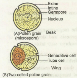
Each pollen grain (microspore) is unicellular, uninucleate, spheical or oval haploid structure.
It is with a double layered wall called sporoderm.
The outer layer of pollen wall is thick, highly resistant and is called exine.
The exine may be spiny (as in insect pollinated plants) or smooth (in wind pollinated plants).
Exine is mainly composed of a complex substance called sporopoiienin which provides resistance to a pollen grain from physical and biological decomposition.
Exine is interrupted at one or more places, which look like small pores called germ pores.
Pollen grains are mostly uniporate (single germ pore) in monocots and triporate (three germ pores) in dicots.
The inner layer of sporoderm is called intine; it is composed of cellulose and pectin. It encloses protoplasm with single haploid nucleus.
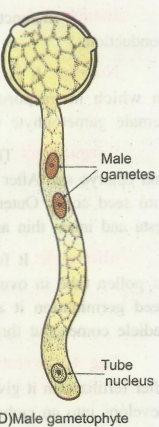
Development of male gametophyte
Microspore or pollen grain is the initial cell of male gametophyte.
The development of male gametophyte is endosporic i.e. occurs within the microspore.
It involves only two mitotic divisions. It is completed in two stages at two different places; before pollination in the pollen sac and after pollination on the stigma.
Before Pollination in the pollen sac:
While in-situ (i.e. when it is still enclosed within pollen sac) the protoplast of pollen grain divide mitotically (by mitosis) to from two unequal cells.
The smaller cell is called generative cell. It has a large nucleus, thin cytoplasm and it lacks reserve food and vacuole.
The larger cell is called vegetative or tube cell, it has a large vacuole, cytoplasm, nucleus and reserve food.
Generative cell lacks a definite cell wall and is freely suspended in cytoplasm of vegetative cell.
In most of the Angiosperms, pollen grains are released at two celled stage after dehiscence of anther.
Such 2- celled pollen grain is also called young or partially developed male gametophyte.
In some Angiosperms, the generative cell divides by mitosis to form two male gametes and thereore, 3-celled pollen grains are released from anther.
After pollination on the stigma:
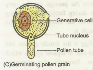
After pollination, 2- celled pollen grain is deposited on stigma surface, comes in contact with sugary stigmatic secretions and absorbs it.
Due to this the volume of cytoplasm increases and creates a pressure which acts on the intine.
The intine of pollen grain comes out of the germ pore in the form of a tube called pollen-tube.
The tube nucleus, cytoplasm and generative cell, all migrate into the pollen tube.
The pollen tube grows down towards ovule through the style due to chemical stimulus inside ovary.
The generative cell divides by mitosis in the pollen tube to form two haploid, non motile male gametes.
This pollen tube with two male gametes, thin cytoplasm and a degenerating sterile vegetative nucleus represents male gametophyte.
Thus it is highly reduced structure.
