Structure of Anatropous Ovule
Gynoecium is the female reproductive floral whorl having individual members called carpels.
Each carpel consists of ovary, style and stigma.
Ovary encloses one or more ovules in it.
An ovule is the integumented megasporangium of the seed bearing plants.
An ovule which has a bent axis and downwardly directed opening called micropyle is termed as anatropous ovule.
It is most common type of ovule in Angiosperms.
Megasporogenesis i.e. formation of megaspore and development of female gametophyte takes place inside the ovule.
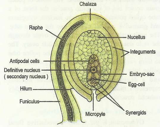
V.S. of mature ovule
It shows following structure: It consists of two main parts, stalk and body.

Stalk:
Stalk of ovule is called funicle or funiculus. It attaches an ovule with the fertile tissue of ovary called placenta.
It has vascular strand and supplies nutrition to the body of ovule.
In anatropous ovule, a considerable part of funicle remains attached with the body.
This pat may even persist in mature seeds and is called raphe.
Body:
The funicle bears a major swollen part of ovule at its tip called body of the ovule.
The stalk/ funicle is attached to the basal part of the body of ovule at a point called hilum.
The body of an ovule consists of following parts;
Nucellus:
It forms the main central bulk of ovule's body.
The nucellus represents the megasporangium proper of an ovule.
It consists of many diploid parenchyma cells.
Chalaza:
The basal part of nucellus from where the integuments develop is called chalaza.
This end of ovule is called chalazal end.
Integuments:
These are the protective coverings of nucellus, which develop from the chalazal part of nucellus and surround it completely except a small portion at the opposite or terminal end.
There are two integuments, outer and inner and therefore, ovule is called bitegmic.
Angiospermic ovules are generally bitegmic.
Micropyle:
The integuments leave a narrow opening at the terminal end of nucellus.
It is called micropyle. This end of nucellus is called micropylar end.
Embryo Sac:
In a mature ovule, nucellus shows the presence of an oval shaped, haploid, structure at micropylar end, this is called embryo sac or female gametophyte.
It consists of 7 cells and 8 nuclei.
There is a 3- celled egg apparatus at the micropylar end.It consists of central egg cell (oosphere) and two lateral cells called synergids.
At the chalazal end, it has three antipodals.
A single large central cell which is covered with mother wall of megaspore, it has two haploid polar nuclei at the centre.
These polar nuclei fuse with each other at the later stage (before fertilization) to form diploid secondary nucleus.
Generally, embryo sac in Angiosperms is monosporic, endosporic, 7-celled and 8 nucleate. It is called Polygonum type.
Functions of different parts of Ovule
Funicle:
It functions for support, projection and conduction.
Nucellus:
It is the megasporangium of ovule, in which megasporogenesis and development of female gametophyte (embryo sac) takes place.
Integuments:
They give protection to nucellus and embryo sac.
After fertilization, these are converted into seed coats. Outer, thick and resistant is called testa and inner, thin and transpaent is called tegmen.
Micropyle:
It forms the passage for the entry of pollen tube in ovule during fertilization. During seed germination it allows the entry of water and radicle comes out through it.
Egg Apparatus:
Egg is the female gamete and after fetilization it gives ise to diploid zygote which develops into an embryo.
Synergids play supportive role in fertilization and degenerate after fertilization.
The filiform apparatus of synergids attracts pollen tube duing fertilization.
Polar Nuclei:
Both the polar nuclei fuse with the second male gamete and form primary endosperm nucleus.
It develops into a nutritive tissue called endosperm (3n). It nourishes the developing embryo.
Antipodals:
These are accessory cells, which degenerate after fertilization.
Development of Female gametophyte
A diploid hypodermal cell at the micropylar end of nucellus gets differentiated to form archespoium.
Mostly this single-celled archesporium directly functions as megaspore mother cell (MMC).
The diploid MMC (2n) undergoes meiosis to form a tetrad of haploid megaspores (n).
This process is known as megasporogenesis Megaspores are generally arranged in linear tetrad.
Generally the chalazal megaspore remains functional while remaining three degenerate gradually.

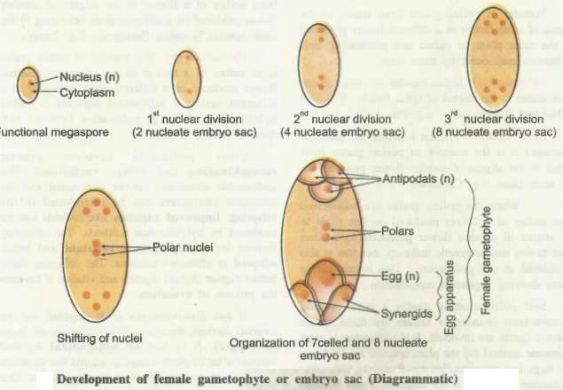
Functional (fertile) megaspore is the first cell of female gametophyte. It undergoes enlargement and develops into a female gametophyte.
The haploid nucleus of functional megaspore undergoes three successive free-nuclear mitotic divisions.
First mitotic division results in formation of two nuclei. Both the nuclei undergo two successive divisions.
This results in formation of four nuclei at each pole and an 8-nucleated structure is formed.
One nucleus from each pole comes to the center and they function as polar nuclei.
This is followed by cellular organization to form 3-celled egg apparatus at micropylar end, three antipodals at chalazal end and two polar nuclei remain in the centre.
Thus, 8-nucleated, 7-celled female gametophyte is formed within the functional megaspore; therefore the development is called endosporic.
Only one megaspore takes part in the formation of embryo sac; therefore it is called monosporic. (In some Angiosperms, embryo sac may be bisporic or tetrasporic)

POLLINATION: TYPES AND AGENCIES:
The process of transfer of pollen grains from anther to the stigma of flower is described as pollination.
It is an initial and essential step of fertilization process in Angiosperms and all seed bearing plants.
Process of pollination was first discovered by Camerarius: (1694).
He described pollination as an essential process for the formation of seeds.
